Cone beam computed tomography (CBCT)
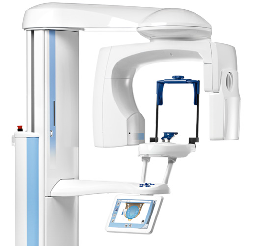
Cone beam computed tomography (CBCT) is a radiographic imaging technique consisting of X-ray computed tomography where the X-rays are divergent, forming a cone. It is required when 2 dimensional xray is not enough to diagnose your problem or plan your treatment.
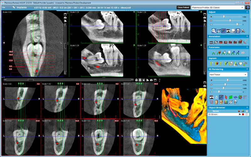
This technology is used to produce three dimensional (3-D) images of your teeth, soft tissues, nerve pathways and bone in a single scan.

CBCT scanners have many uses in dentistry:
- oral and facial surgery
- surgical planning for impacted teeth.
- diagnosing temporomandibular joint disorder (TMJ).
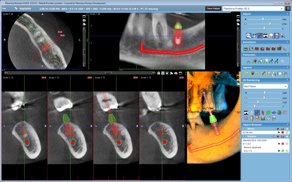
- accurate placement of dental implants.
- evaluation of the jaw, sinuses, nerve canals and nasal cavity.
- detecting, measuring and treating jaw tumors.
- determining bone structure and tooth orientation.
- locating the origin of pain or pathology.
- cephalometric analysis.
- reconstructive surgery
- root canal treatment for locating missed canals.

During the scan, the CBCT scanner rotates around the patient’s head, obtaining up to nearly 600 distinct images. It is not a claustrophobic sensation like CT SCAN and the patient is standing or sitting for a very small duration of time. The scanning software collects the data and reconstructs it, producing three dimensional images of your mouth. These images can then be manipulated and visualized with specialized software. The radiation exposure is more than a regular xray and less than a CT Scan.

